Cancer metabolites organize the tumor microenvironment

Cancer cells are not alone: As cancerous tumors grow unregulated cancer cells engage other cells, in their path of destruction like macrophages which are part of the immune system and endothelial cells which make blood vessels. This collection of non-cancer cells that powers cancer growth is called the tumor microenvironment. How does the tumor microenvironment help cancer cells proliferate even more?

Answering this question is a holy grail of cancer science and holds the key to new therapies. Cancer cells, with their many mutations and unchecked DNA damage, change constantly: they can be a moving target for therapy and develop resistance to drugs that seemed to work at first. The non-cancer cells in the tumor microenvironment are genetically stable. If we knew how these cells interact we could to stop the tumor microenvironment from feeding the cancer, halt cancer growth or even reverse it. On February 28 a team of SKI scientists published a significant advance. The answer—surprisingly—is in metabolism.

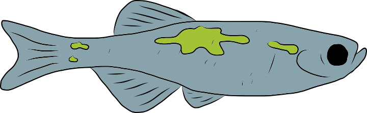
All cells rely on metabolism, the engine-like process that requires constant fuel and oxygen to run. Cancer cells have altered metabolisms: they consume lots of oxygen and dump metabolic waste such as lactic acid. Because of this, cancerous tumors should only grow so large before the toxic effects accumulate like pollution in a jam-packed city, and eventually slow cancer growth. This is prevented, however, by tumor-associated macrophages (TAMs) that respond to the harsh environment and start a tissue-repair mechanism to clean it up.

The SKI team started by observing the behaviors of TAMs in a mouse model of cancer. Then, they fabricated tissue-mimetic systems to recreate the same process in vitro. Using this approach they discovered that TAMs respond to low oxygen and to the presence of lactic acid and start producing a vascular endothelial growth factor (VEGF). This growth factor commands endothelial cells to start producing blood vessels—called neo-angiogenesis—a process that can bring new blood to struggling cancer cells, replenishing oxygen and removing toxic waste.

The tissue-repair response of macrophages is normally a good thing: it is how our body heals wounds and clears out toxic waste from muscles after intense exercise. In cancers, however, it can make cells with complementary skills—cancer cells, TAMs and endothelial cells—work together in a terrible way. Rescuing cancer cells from dying because of their own altered metabolism boosts the cancer to grow even more.

The SKI study established a new role for cancer metabolism in the interactions between cancer cells and their microenvironment. These findings lay the foundations for our understanding of cancer development, diagnosis and treatment.

But the study also showed how an interacting team of multidisciplinary scientists could answer a difficult cancer question: Craig Thompson brought his expertise in cancer metabolism, Johanna Joyce her expertise in the tumor microenvironment, and João Xavier his expertise in cancer systems biology, a new field that aims to integrate cancer concepts. Cancers subvert cells with complementary features in their path to destruction; figuring out its complex mechanisms—and new ways to fight them—may require teams of scientist with complementary skills.

The study was spearheaded by Carlos Carmona Fontaine, former postdoctoral researcher at MSKCC who is now assistant professor of Biology at the New York University.

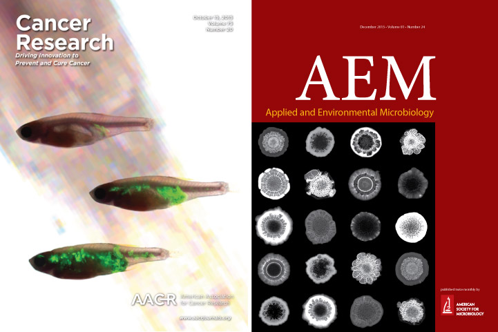
Metabolic origins of spatial organization in the tumor microenvironment
Carlos Carmona-Fontaine, Maxime Deforet, Leila Akkari, Craig B. Thompson, Johanna A. Joyce, Joao B. Xavier. PNAS
[Open Access]

Posted in Uncategorized | Leave a comment

2016 lab mug

mug2016Carlos Carmona Fontaine, in between finishing his paper and starting his lab at NYU, found time to make the official 2016 Xavier Lab MUG.

Colored in the beautiful parula map, the 2016 mug is the best mug in the world for coffee or tea.

Check out Carlos’s lab website carmofon.org. Join his new lab at NYU Biology and you might make it in time for the first-ever Carmona Fontaine Official Mug.

Read more about the 2016 lab mug and our other mugs in our paraphernalia page.

Posted in Uncategorized | Leave a comment

NY/B.I.G. meeting

This Friday 10:00am – 6:15pm at NYU
Image

Kerry Boyle talks about “The metabolomic basis of social behavior” at 10:20-10:35.
More about it: http://biology.as.nyu.edu/object/biology.events.nybig2016

Posted in Uncategorized | Leave a comment

High school students and teachers at MSK for the 2015 “Major Trends in Modern Cancer Research”

On Tuesday, November 10, MSK hosted the tenth annual “Major Trends in Modern Cancer Research” lecture for high school and college students. The session was moderated by Craig Thompsons and featured Kat Hadjantonakis, Cole Hanes and me. The whole session is on youtube.

High school students at MSK for the 2015

High school students at MSK for the 2015 “Major Trends in Modern Cancer Research”

Posted in Uncategorized | Leave a comment

Journal covers

During my PhD, 1999-2003, most scientific articles I needed for my research where only available in print. I’d go down to the library and take hours photocopying articles from piles of journals.
I also listened to a lot of CDs and I loved the cover art. I haven’t picked up a CD in a while and I’m pretty sure new CDs are still made, but I don’t know who buys them. The art from new covers goes unnoticed – I haven’t seen one from the last 5 years or more.
Journals still get printed too, but like songs we get the articles online. Grad students don’t spend hours copying in the library, which is great. But some nice covers will go unnoticed.

Covers from the October 15 issue of Cancer Research (collaboration with Richard White's lab) and the December 2015 about issue of Applied and Environmental Microbiology (collaboration with Lars Dietrich's lab and Soren Sorens's lab).

Covers from the October 15 issue of Cancer Research (collaboration with Richard White’s lab) and the December 2015 issue of Applied and Environmental Microbiology (collaboration with Lars Dietrich’s lab and Soren Sorensen’s lab).

Posted in Uncategorized | Leave a comment

Systems Biology talk at the MSKCC presidential seminars this week

Evolutionary Tradeoffs and the Geometry of Gene Expression Space
Uri Alon, PhD
Senior Scientist, Department of Molecular Cell Biology and Department Physics of Complex Systems
Weizmann Institute of Science
Rehovot, Israel

October 21, 2015 at 4:30 PM
Host: GSK Graduate Students
ZRC Auditorium

This talk is followed by a special seminar “The Emotional Sides of Science: a Guitar Talk”

Posted in Uncategorized | Leave a comment

Taking statistical pictures of metastasis using zebrafish

Metastasis, the spread of cancer from its primary site to other parts of the body, is when cancer gets really bad. But this process is poorly understood, in large part because it is stochastic. Like the dispersal of plant seeds across a fertile field, metastatic cells could end up in many different places so knowing where metastases will form exactly may seem impossible.
What we need to study physical phenomena that are stochastic is lots of samples and statistics. Studying metastasis across large samples can be very difficult however. Investigating patterns of metastasis in the human body would require compiling dozens of cases while controlling for factors that could influence metastasis in unknown ways like patient age, body-mass-index, exposure to carcinogens, etc. We could use animal models and control for these factors, but common models in cancer research like mice and rats are still quite expensive to run experiments with dozens of samples.

“The zebrafish” (2015) by Silja Heilmann

Enter the glorious zebrafish. The zebrafish is already a powerful model for genetics and development and it is gaining increasing importance in cancer biology. Our lab collaborates with the lab of Richard White in the program for Cancer Biology and Genetics to investigate metastatic spread across dozens of zebrafish. For the past three years Silja Heilmann has been working closely with Rich, Kajan and other members of the While lab to develop protocols and methods for the quantitative analysis of metastasis. The model is a transparent zebrafish called Casper that is great for imaging. In Rich’s lab, they developed a zebrafish melanoma cell line called Zmel1 that expresses GFP. Once injected into adult casper zebrafish, Zmel1 forms primary tumors that later on produce metastasis and we can visualize the process using microscopy.
Silja developed image analysis algorithms that resize and align many pictures of fish together. This procedure allows building a statistical picture of metastatic growth across the whole animal. The detailed picture reveals indeed the strong stochastic nature of metastatic spread, but some patterns start to emerge. Advancements such as these may one day enable a better understanding of metastasis and help in the development of anti-metastasis treatments.

Read the paper:

A quantitative system for studying metastasis using transparent zebrafish
Silja Heilmann, Kajan Ratnakumar, Erin Langdon, Emily Kansler, Isabella Kim, Nathaniel R Campbell, Elizabeth Perry, Amy McMahon, Charles Kaufman, Ellen van Rooijen, William Lee, Christine Iacobuzio-Donahue, Richard Hynes, Leonard Zon, Joao Xavier, and Richard M White. Cancer Research
[PDF]

Posted in Uncategorized | Leave a comment

Lessons from a frinkandel-shaped cell

Organisms can vary a lot in body size among species, but within species they are very similar

Organisms can vary a lot in body size among species, but within species they are very similar

Organisms of different species have different shapes and sizes and still, within the same species, shape and size are practically constant. For example, elephants can be 250,000 larger in volume than mice, yet when we compare elephants of the same species and controlling for age they shouldn’t  differ by more than 2x (I’m is guessing – I couldn’t find any reference for this). How do organisms within a species maintain relatively constant shapes and sizes when it’s at least physically possible to have wide variations?
This seems a question for developmental biologists, but microbial cell biologists are very interested in their version of the same problem. Bacterial species can vary enormously in cell shape and size. One of the largest, Thiomargarita namibiensis (100–300 µm), can be 1000x longer than one of the smallest known Mycoplasma genitalium (~300 nm). Nonetheless, within a species, their sizes are often constant and, we imply, very well regulated. There was a recent burst of papers where different groups across the US measured with unprecedented precision the distribution of cell lengths in the rod shaped bacteria E. coli, Calaubacter crescentus and Bacilus subtilis. From this collective work a simple mechanism seems to emerge: Rod shaped bacteria decide when to divide by determing that they added a fixed increment to their cell length. This so called “incremental rule” (a.k.a. “adder model” or “constant size extension“) had been proposed earlier but lacked empirical support. It provides a simple explanation for why cell populations maintain a narrow size distribution – I strongly reccomend reading those papers.
We added additional evidence for the incremental model by showing that the pathogen Pseudomonas aeruginosa, our model organism, also follows the incremental rule. Our interest in this problem came by accident when we isolated a mutant with abnormally long cell from our hyperswarming experiment. In our new paper we analyze the length distribution in wild-type Pseudomonas aeruginosa and in this mutant which we call frik, not only because it is a freak but also because it reminds us of the tasty dutch snack frinkandel.

frik cell on the left and the dutch frikandel on the right. Remarkable similarity.

frik cell on the left and the dutch frikandel on the right: A striking resemblance.

The frikandel is a meat based rod of mysterious composition. It’s probably made with left over meat, but it’s hard to tell exactly what its composition is from it’s spicy taste and spongy texture (hmmm…). Similarly, exactly why the frik cell is abnormally shaped is somewhat a mystery to us. We resequenced the genome of the frik mutant and we found two candidate mutations. We were capable of ruling out one of those two using microbial genetics. The second one, however, is really hard to clone and we could not confirm nor refute that it is the cause of the frik phenotype. One explanation is that the mutation is in a gene (PA14_65570) that may be essential which would make it harder to clone.
Still, our paper extends the reach of the elegant incremental model by showing that it also applies to P. aeruginosa, a common cause of opportunistic infections. We also show that the frik cells are more sensitive to some antibiotics, suggesting that affecting cell size regulation could have implications for improved antimicrobial therapy.

Read our paper:
Cell size homeostasis and the incremental rule in a bacterial pathogen
Maxime Deforet*, Dave van Ditmarsch* and João B. Xavier. Biophysical Journal
[PDF]

Posted in Uncategorized | Leave a comment

When do bacteria decide to cooperate?

Bacteria don’t think since they’re just single celled organisms. But they do make decisions. Take the lac operon, arguably the best studied molecular system that enables an organism to adapt to changing conditions. When bacteria have plenty of a nutrient that they like, glucose, they don’t express the genes to eat up another nutrient that they like less, lactose. When glucose runs out, glucose starvation sends a signal as a series of molecular processes inside the cell that end in the expression of the genes in the lac operon. The enzymes encoded in those genes help the cell break down lactose into glucose and galactose and the cell is happy again.

The lac operon is a cellular decision-making system, but one that is pretty individualistic. When a cell makes a decision to express genes based on whether it lacks a nutrient, the decision affects that cell first and foremost (even though decisions to eat new sugars from the environment can always also affect others that compete for the same food). But bacteria can make decisions that are more social.

Mutants that don't produce surfactants (rhlA- here in green) swarm using the surfactants produced by wilt-type bacteria (in red)

Mutants that don’t produce surfactants (rhlA- here in green) swarm using the surfactants produced by wilt-type bacteria (in red)

In our lab we study how cells make decisions to cooperate with other bacteria. Bacteria have many collective traits like biofilms and swarming, which can only happen when there are many cells in a community. One thing in common for these traits to happen is that many individual bacteria need to come together and start producing substances that accumulate in the extracellular space. To build a biofilm, bacteria must secrete large amounts of polymers and make an extracellular matrix. In order to swarm, bacteria must secrete lots of rhamnolipid surfactants and lubricate the surface of a Petri dish. Each cell must spend resources to produce their individual share of a common good. Without these individual contributions the social trait could happen. Cheaters within the population (like the rhlA- strain in this picture) could take advantage of the public good without themselves contributing.

P. aeruginosa decides to cooperate only when they are in a crowd (quorum sensing) and have enough carbon source due to growth limitation by some other nutrient (iron, in this case)

P. aeruginosa decides to cooperate only when they are in a crowd (quorum sensing) and have enough carbon source due to growth limitation by some other nutrient (iron, in this case)

How can cooperating cells prevent cheating? One way is to invest in cooperation only when there are plenty of resources to do so. We uses quantitative experiments and mathematical modeling to analyze how bacteria do this. We saw that bacteria decide to express genes to make the surfactants needed to swarm when they have more than enough carbon rich nutrients, a process that we call metabolic prudence. However, they also count their neighbors using a process called quorum sensing. This way, each individual bacterium checks if it has enough nutrients and enough neighbors for swarming before cooperating. At any point in this process, if the bacterium gets starved too severely it shuts down cooperation, possibly to dedicate resources in preparing for survival.

Check out our paper, out this week in PLoS Computational Biology.

Integration of Metabolic and Quorum Sensing Signals Governing the Decision to Cooperate in a Bacterial Social Trait
Kerry E. Boyle, Hilary Monaco, Dave van Ditmarsch, Maxime Deforet, Joao B. Xavier

Posted in Uncategorized | Leave a comment

2015 lab mug

Here’s the 2015 lab MUG designed by Silja Heilmann. Great for warm coffee or tea on a cloudy New York Sunday.

labmug2015

Posted in Uncategorized | Leave a comment